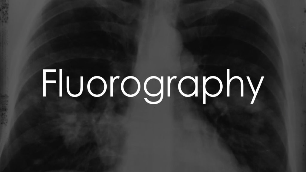
Fluorography, also known as chest fluorography, is a diagnostic imaging technique that uses X-rays to produce images of the chest area, particularly the lungs. This method is widely used for screening and diagnosing various pulmonary conditions, including tuberculosis (TB), pneumonia, lung cancer, and other respiratory diseases. Fluorography is valued for its ability to detect abnormalities in the lungs early, allowing for timely intervention and treatment. In this article, we will explore the purpose of fluorography, the procedure for conducting it, the interpretation of results, and important considerations when undergoing this test.

Why is it needed?
Fluorography is necessary for several critical reasons:
- Screening for Tuberculosis: It is commonly used in public health programs to screen for TB, especially in high-risk populations.
- Diagnosing Lung Diseases: Helps in diagnosing conditions such as pneumonia, bronchitis, and lung cancer.
- Monitoring Chronic Respiratory Conditions: Used to monitor diseases like chronic obstructive pulmonary disease (COPD) and pulmonary fibrosis.
- Preoperative Assessment: Evaluates lung health before surgery, particularly in patients with a history of respiratory issues.
- Workplace Health Monitoring: Regular screening for workers exposed to respiratory hazards, such as silica or asbestos.
Procedure for taking the test
The procedure for conducting fluorography involves several steps:
Preparation
- Clothing: Patients may be asked to remove clothing from the upper body and wear a hospital gown.
- Jewelry and Accessories: Remove any metal objects, including necklaces, earrings, and body piercings, as they can interfere with the X-ray images.
- Breathing Instructions: Patients are usually instructed to take a deep breath and hold it during the imaging to get a clear picture of the lungs.
Positioning
- The patient stands in front of the fluorography machine with their chest against the image plate.
- The arms are usually positioned at the sides or lifted above the head to avoid obstructing the view.
Image Acquisition
- The technician operates the machine from behind a protective barrier to avoid radiation exposure.
- A quick X-ray image is taken, typically lasting just a few seconds.
- The entire process is generally quick and painless.
Completion
- After the image is taken, the patient can usually get dressed and resume normal activities immediately.
Interpretation of results
Interpreting fluorography results involves examining the X-ray images for any abnormalities in the lungs and surrounding structures. Here are some key findings and their potential implications:
Normal Findings
- Clear Lungs: No signs of infection, masses, or fluid accumulation.
- Normal Heart Size: The heart is of a normal size and shape.
- Unobstructed Airways: No blockages or narrowing in the airways.
Abnormal Findings
- Infiltrates: Areas of increased density, often indicating pneumonia, tuberculosis, or other infections.
- Nodules or Masses: Could suggest lung cancer, benign tumors, or granulomas.
- Pleural Effusion: Fluid accumulation between the layers of tissue lining the lungs, possibly due to infection, heart failure, or malignancy.
- Enlarged Heart: May indicate heart conditions such as cardiomegaly or heart failure.
- Fibrosis: Scarring of lung tissue, commonly seen in conditions like pulmonary fibrosis.
- Atelectasis: Collapse of part of the lung, often due to blockage or compression.
Important Considerations
When undergoing fluorography, several factors should be taken into account:
- Radiation Exposure: Although the radiation dose from fluorography is low, it is still important to limit unnecessary exposure. Inform the technician if you are pregnant or suspect you might be.
- Follow-Up Tests: Abnormal findings may require further diagnostic tests, such as a CT scan, MRI, or biopsy, to confirm the diagnosis and determine the appropriate treatment.
- Symptom Correlation: Results should be interpreted in conjunction with clinical symptoms and medical history for accurate diagnosis and management.
- Regular Screening: For individuals at high risk of lung diseases, regular fluorography screening may be recommended as part of a routine health check-up.
Conclusion
Fluorography is a valuable diagnostic tool that provides critical information about the health of the lungs and surrounding structures. It is a quick, non-invasive, and effective method for screening and diagnosing various pulmonary conditions. Understanding the purpose of the test, the procedure involved, and the interpretation of results can help patients better prepare for the examination and understand the findings. Regular fluorography, particularly for individuals at high risk for respiratory diseases, can significantly aid in early detection and effective management of lung conditions.