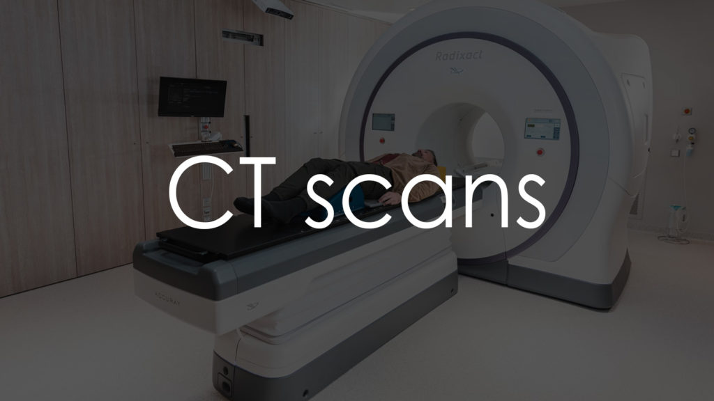
A computed tomography (CT) scan, also known as a CAT scan, is a non-invasive imaging test that combines X-ray measurements taken from different angles to create cross-sectional images of the body. This advanced diagnostic tool provides more detailed information than standard X-rays, allowing for a more precise evaluation of internal organs, bones, soft tissues, and blood vessels. CT scans are essential for diagnosing and monitoring a wide range of medical conditions, guiding treatment plans, and performing interventional procedures. This article will explore the purpose of a CT scan, the procedure for conducting it, the interpretation of results, and important considerations when undergoing this test.

Why is it needed?
A CT scan is necessary for several critical reasons:
- Diagnosing Medical Conditions: It helps identify conditions such as tumors, infections, blood clots, and fractures.
- Evaluating Symptoms: It is used to investigate symptoms like severe pain, unexplained weight loss, and persistent headaches.
- Monitoring Chronic Conditions: Regular CT scans can monitor the progression of diseases like cancer, cardiovascular disease, and inflammatory conditions.
- Guiding Procedures: It aids in guiding biopsies, needle aspirations, and other minimally invasive procedures.
- Assessing Trauma: It is commonly used in emergency settings to assess injuries from accidents or trauma.
Procedure for taking the test
The procedure for conducting a CT scan involves several steps:
Preparation
- Clothing: Patients may need to change into a hospital gown, depending on the area being examined.
- Jewelry and Accessories: Remove any metal objects, including jewelry, glasses, and body piercings, as they can interfere with the imaging.
- Contrast Material: Some CT scans require the use of a contrast material to enhance the visibility of certain structures. This can be administered orally, intravenously, or rectally. Patients may be asked to fast for a few hours before the scan if contrast material is used.
Positioning
- Lying Down: The patient lies on a motorized table that slides into the CT scanner’s circular opening.
- Positioning the Body: The technologist positions the body part to be imaged at the center of the scanner. Straps and pillows may be used to help maintain the correct position and keep the patient still.
Image Acquisition
- Taking the Scan: The CT scanner rotates around the patient, taking X-ray measurements from various angles. These measurements are processed by a computer to create cross-sectional images (slices) of the body.
- Holding Still: Patients may be asked to hold their breath for a few seconds during the scan to avoid blurring the images.
- Duration: The scan itself typically takes just a few minutes, but the entire appointment, including preparation, may take about 30 minutes to an hour.
Completion
- Reviewing the Images: The technologist checks the images to ensure they are clear and complete.
- Final Steps: Once the images are satisfactory, the patient can usually resume normal activities immediately. If contrast material was used, instructions for post-scan care may be provided.
Decoding the results
Interpreting CT scan results involves examining the cross-sectional images for abnormalities in the bones, organs, and other structures. Here are some key findings and their potential implications:
Bones
- Fractures: Visible breaks or cracks in the bone.
- Bone Density: Changes in density can indicate conditions like osteoporosis or bone tumors.
- Deformities: Abnormal shapes or misalignments in bones.
Brain
- Hemorrhages: Areas of bleeding within the brain.
- Tumors: Abnormal masses or growths.
- Stroke: Areas of reduced blood flow indicating ischemic stroke or bleeding indicating hemorrhagic stroke.
- Atrophy: Reduction in brain tissue volume, which can be associated with neurodegenerative conditions.
Chest
- Lung Conditions: Detection of tumors, pneumonia, pulmonary embolisms, or interstitial lung disease.
- Heart and Vessels: Evaluation of heart size, aortic aneurysms, and other vascular abnormalities.
Abdomen and Pelvis
- Organ Assessment: Detailed images of the liver, kidneys, pancreas, spleen, and other organs to identify tumors, cysts, infections, and inflammations.
- Bowel Conditions: Detection of obstructions, perforations, diverticulitis, or inflammatory bowel disease.
- Urogenital System: Evaluation of the bladder, prostate, ovaries, and uterus for tumors, cysts, or other abnormalities.
Vascular System
- Aneurysms: Abnormal bulging in blood vessels.
- Blockages: Detection of blood clots or plaque buildup in arteries.
- Dissections: Splitting of the arterial wall layers, which can be life-threatening.
Important Considerations
When undergoing a CT scan, several factors should be taken into account:
- Radiation Exposure: Although the radiation dose from a CT scan is higher than that from a standard X-ray, it is generally low and considered safe. However, unnecessary exposure should be avoided.
- Pregnancy: Inform the technologist if you are pregnant or suspect you might be, as radiation can be harmful to the developing fetus.
- Allergies: Inform the healthcare provider about any known allergies, especially to contrast material, as it can cause allergic reactions in some individuals.
- Hydration: If contrast material is used, staying well-hydrated before and after the scan can help flush the contrast out of the body.
- Follow-Up Tests: Abnormal findings may require additional imaging tests, such as MRI or ultrasound, for further evaluation and confirmation.
- Symptom Correlation: Results should be interpreted in conjunction with clinical symptoms and medical history for accurate diagnosis and management.
Conclusion
A CT scan is a vital diagnostic tool that provides detailed cross-sectional images of the body’s internal structures. It is non-invasive, quick, and widely accessible, making it an essential procedure in modern medicine. Understanding the purpose of the test, the procedure involved, and the interpretation of results can help patients better prepare for the examination and understand the findings. Regular CT scans, particularly for individuals with symptoms or risk factors for certain conditions, can significantly aid in early diagnosis and effective management of various medical issues.